Identifying the 19 types of spots on your skin requires distinguishing between benign growths and conditions that necessitate professional clinical evaluation. According to the American Academy of Dermatology (AAD), skin lesions—ranging from red, pink, and brown marks to various textures—often stem from sun exposure, genetics, or common inflammatory responses. While many skin spots are harmless, dermatologists emphasize that any lesion changing in size, shape, or color should be examined to rule out malignancy.
As a physician, I frequently see patients concerned about “new bumps” or discolored patches. While it is easy to worry, most skin changes are common dermatological conditions. However, the National Cancer Institute notes that early detection of atypical growths is the most effective strategy for managing skin health. Understanding the visual characteristics of these spots is the first step toward effective communication with your healthcare provider.
Common Brown and Darkly Pigmented Skin Lesions
Brown spots are often the result of concentrated melanin. The most common of these include solar lentigines, frequently called “age spots,” which appear due to long-term ultraviolet (UV) exposure. According to the Mayo Clinic, these are benign but serve as a clinical marker for cumulative sun damage. Another frequent finding is the seborrheic keratosis, which often has a waxy, “pasted-on” appearance and can range from light tan to deep black.
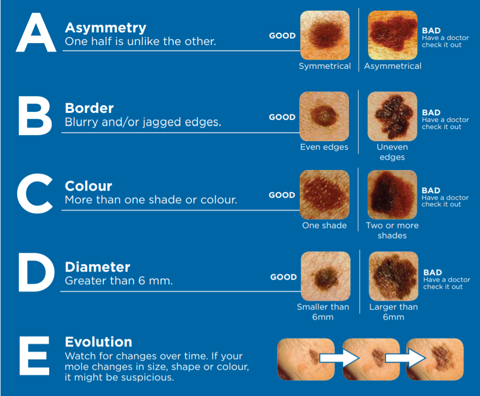
Dermatologists also monitor for nevi, commonly known as moles. While most moles are benign, the Skin Cancer Foundation advises patients to use the “ABCDE” rule—looking for Asymmetry, Border irregularity, Color variation, Diameter larger than a pencil eraser, and Evolving features—to identify suspicious growths. Freckles (ephelides) also fall into the brown category, typically darkening after sun exposure and fading in the winter months.
Red and Pink Spots: Inflammation and Vascular Conditions
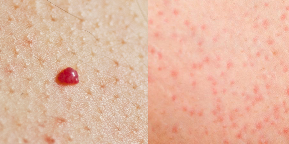
Red or pink spots frequently signal an inflammatory response or vascular dilation rather than pigmentation issues. One common example is cherry angiomas, which are small, bright red papules composed of clusters of capillaries. These are generally harmless and tend to increase in number with age. Conversely, persistent red, scaly patches might indicate actinic keratosis, which the American Academy of Dermatology classifies as a precancerous lesion caused by chronic sun damage.
Other common red or pink spots include:
- Dermatofibromas: Firm, reddish-brown bumps that often feel like a hard knot under the skin.
- Pyogenic Granulomas: Rapidly growing, bright red bumps that bleed easily, often appearing after minor trauma.
- Rosacea-related papules: Small, pink bumps often accompanied by facial redness and visible blood vessels.
- Psoriasis plaques: Thick, red patches covered with silvery scales, typically appearing on the elbows, knees, or scalp.
Clinical Indicators for Professional Evaluation
Determining whether a spot requires a biopsy depends on its behavior over time. A “stable” spot—one that has not changed in years—is often benign. However, clinical guidelines from the National Health Service (NHS) in the United Kingdom suggest that any lesion that bleeds, crusts, or fails to heal within four weeks warrants a professional assessment. Relying on self-diagnosis for changing skin lesions is discouraged due to the risk of misidentifying early-stage carcinomas.
When you visit a dermatologist, they will likely use a dermatoscope, a specialized magnifying tool, to examine the subsurface structure of the spot. This allows the clinician to see patterns of pigment or vascularity that are invisible to the naked eye. In cases of clinical uncertainty, a biopsy—the removal of a small sample of tissue for laboratory analysis—remains the gold standard for diagnosis.
Preventative Measures and Ongoing Monitoring
Maintaining skin health involves consistent UV protection. The World Health Organization (WHO) recommends broad-spectrum sunscreen with an SPF of at least 30, protective clothing, and avoiding peak-hour sun exposure to mitigate the development of many types of skin spots. While some lesions are genetic, minimizing UV-induced damage significantly lowers the risk of developing actinic keratoses and certain skin cancers.

If you notice a new or changing spot, do not wait for it to resolve on its own. Schedule an appointment with a board-certified dermatologist for a baseline skin exam. Many health systems now offer teledermatology services for initial screenings, though an in-person physical exam is often required for definitive diagnosis. Please share your experiences or questions in the comments section below, and ensure you keep a record of any changes in your skin’s appearance to discuss at your next medical consultation.